At first, I’d like to express my deep respect for the great efforts of those who involving to the fight against the novel coronavirus disease, COVID-2019.
The worldwide great efforts have been revealing properties of the new coronavirus, 2019-nCoV. At the present time, approx. 4 months after discovery of the virus, it is known that 2019-nCoV is a betacoronavirus which has high genomic similarity with SARS coronavirus (SARS-CoV) and can infect human by binding to angiotensin-converting enzyme 2 (ACE2), the same receptor of SARS-CoV, based on the brief review of Japanese Society of Virology.
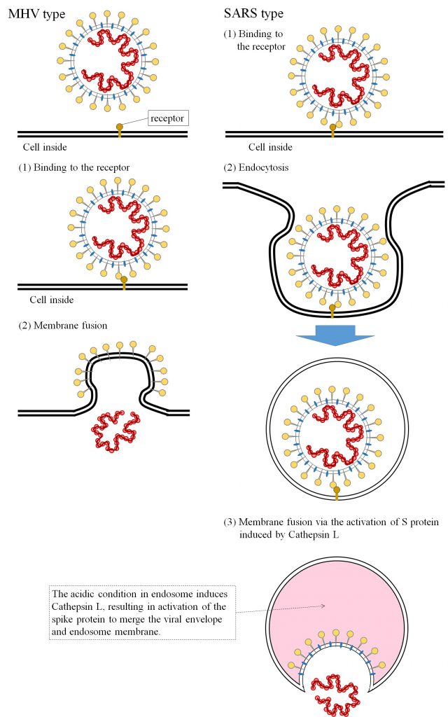
There are two types of cell entry of envelope virus, direct membrane fusion and receptor-mediated endocytosis. As for the mechanism of infection of coronavirus, tremendous investigations has been demonstrated against SARS shock and revealed SARS-CoV can entry into the host cell via the receptor-mediated endocytosis, while the other betacoronavirus, Murine Hepatitis Virus (M-CoV), can deliver its genome into the targeted cell by fusing their membrane with the plasma membrane of host cell directly [736].
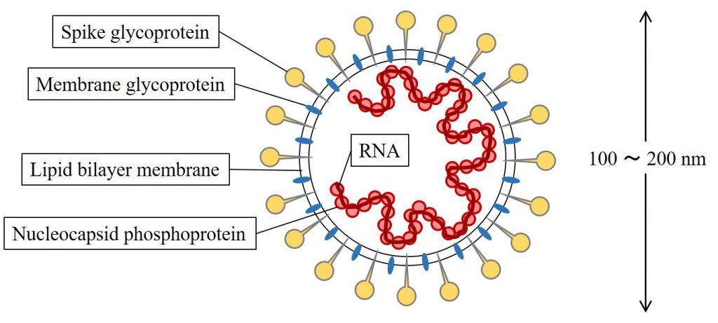
Although there are two types of cell entry of coronaviruses, the spike protein of virion has a key role on the both type of coronavirus infection [736]. As well-known now, the virion structure of coronavirus is schematically drawn like the picture below.

A schematic image of coronavirus, drawn by the author of this post based on the review[736].

A schematic drawing of cell entry mechanism of M-CoV and SARS-CoV, depicted by the author of this post based on the review[736].
On the other hand, the cell entry of SARS-CoV has several steps. At first, SARS-CoV binds to the receptor ACE2. Then the virus can move into the endosome through endocytic pathway. Under the acidic environment of endosome, the endosomal protease “Cathepsin L” activates the spike proteins of virus to induce membrane fusion between envelope and endosome, resulting in the release of viral RNA into the host cell.
There are some treatments of viral diseases, vaccines to train our immune system against the target pathogen, administration of interferons for inhibition of viral replication and the improvement of lifestyle by heathy diet and optimal sleep.
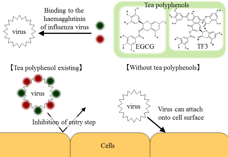
In addition to these treatments, many reports and reviews indicate that tea polyphenols are effective for various diseases accounting for envelope viruses such as influenza [312,750,751,753,754], herpes [308], HIV [310] etc., as posted before. And some of them imply tea polyphenols can adsorb on the surface of envelope viruses, resulting in the decline of infectivity of each envelope viruses. For example, Nakayama et al. (1993) investigated epigalloctechin gallate (EGCG) and theflavin digallate (TF3) can inhibit the infectivity of influenz A and B viruses in Madin-Darby dcanine kidney cells in vitro due to agglutination of influenza virus and tea polyphenols like antibody[312].

A schematic diagram of the mechanism that tea polyphenols prevent influenza virus from infecting, suggested by Nakayama M. et al. (1993) [312].
However, Hubei province, to which Wuhan city belongs, is known as a major tea exporting and consuming area in China. Why tea drinking cannot prevent 2019-nCoV from infecting the people in Wuhan?
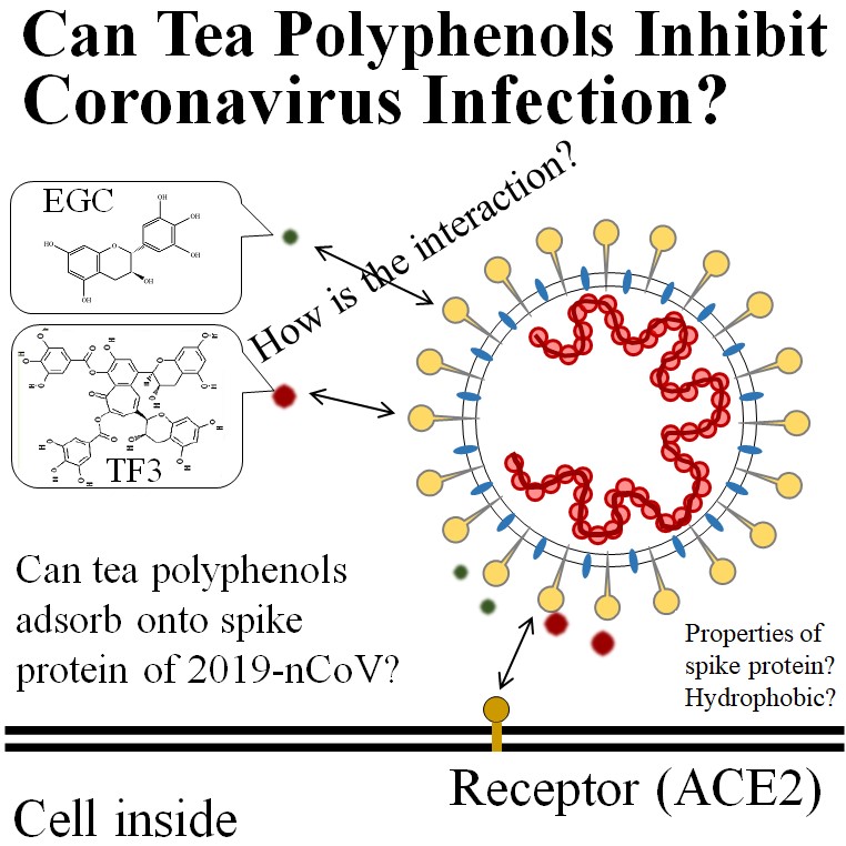
I think further study is necessary to investigate 2 aspects of tea polyphenols for the inhibition of 2019-nCoV infection.
The first one is the interaction between the spike protein of 2019-nCoV and tea polyphenols. The interaction may depend on the type of tea polyphenols, such as EC, ECG, EGC, EGCG, theaflavin and thealubigin etc. It is necessary to investigate the properties of the spike protein of 2019-nCoV in order to analyze the viron-tea polyphenol interaction. If the spike protein has relatively higher hydrophobicity, gallate-type catechins (ECG and EGCG) may prefer to absorb onto the spike protein due to their hydrophobicity around ester binding to galloyl group, based on the report about interaction between tea catechins and biomolecules [747]. On the other hand, tea catechins themselves may not prevent 2019-nCoV infection based on the report that yellow tea produced in Hubei province has higher content of ECG and EGCG [745]. Tea polyphenols of higher molecule weight, such as theaflavins and thealubigins, or complexes of tea polyphenols and other compositions may be more effective for the inhibition of 2019-nCoV infection due to their higher hydrophobicity. On the other hand, the interaction derived from electrostatic properties (or interaction of electric double layer) may suppress the adsorption of tea polyphenols onto the spike protein, if the spike protein has negative charge, based on the result that adsorption of tea catechins on the liposome having relatively high negative charge decreased compared with non-charged liposome [747]. In this case, the composite of positive charged materials and tea polyphenols may be effective. (For example, complexs of tea catechins and chitosan nanoparticle having positive charge. [241])
In addition, further understanding the interactions between tea polyphenols and the spike protein would require the thermodynamic profile of interactions of each tea polyphenol and the spike protein. For this purpose, isothermal titration calorimetry could result in various thermodynamic parameters related to the interactions. Unfortunately I could not search any articles describing the interactions between the spike protein of SARS-CoV and tea polyphenols at the present time, although there may be various reports about the information. Molecular dynamics calculation could be also helpful to estimate the effective materials adsorbing on the spike protein based on the 3 dimensional properties and components of spike protein and target materials. The efforts of Chinese researchers had clarified the genome sequence of 2019-nCoV and the spike protein consists of 1273 amino acids [728]. As reported by global broadcasts such as BBC, various research institutes and biochemical companies has accelerated their investigation using the genomic information. On the early February, Coutard et al. has already reported the sequence of spike protein of 2019-nCoV has a specific furin-like cleavage site, which is absent in the linage b coronaviruses including SARS-CoV [730]. The report would indicate the interaction of the spike protein of COVID-19 with tea polyphenols is different from that of SARS-CoV.
The second one is behavior of tea polyphenols and 2019-nCoV in mucous layer on the respiratory tract. In general, pathogen virions deposit on mucous membrane and upper respiratory tract and then the viruses migrate to the surface of respiratory tract tissue. If tea polyphenols can make 2019-nCoV inactive by adsorption, we should keep tea polyphenols retain in mucous layer as long as possible and interact with spike proteins of virions as efficiently as possible. In order to consider the mechanism of tea polyphenols interacting with virions in mucous layer, it is required to know more the behavior of tea polyphenols and 2019-nCoV in mucus. The transport of virion through mucus to epithelial cells is driven by diffusion [755]. Thus the retention time of virions in mucous layer depends on the size of virion, viscosity of mucous solution based on the diffusion coefficient, thickness of mucous layer and the velocity of advection derived from ciliary movement of periciliary layer. Tea polyphenols also move by diffusion due to thermal motion and advection by ciliary movement. It is ideal that tea polyphenols can retain longer in periciliary layer due to the interaction with cilia than pathogen virions, I think. However, I could not search adequate articles describing interaction of periciliary layer with virions and polyphenols. There are some types of interaction between surfaces of biomaterial, van der Waals interaction, electric double layer, solvation, hydrophobic interaction and hydrogen bond, based on the famous textbook “Intermolecular and Surface Forces” written by Israelachivili. Considering past investigations, the composite of hydrocolloids of naturally occurring materials and tea polyphenol may be effective for the prevention of viral infection. For example, many reports indicate chitosan nanoparticles could be effective and operative to delivery tea polyphenol and promote their intake in gastrointestinal tract due to the longer retention in mucous layer.
Based on the research report by Kao Co. Ltd., the longer retention of tea polyphenols on mucous membrane of throat can be more effective to prevent influenza virus from infecting human. The result shows the oral administration of the mixture of mucin and tea catechins resulted in the decline of influenza infection significantly.
Besides, there are other interesting reports about beneficial effects of tea polyphenols against viral diseases. Monobe et al.(2010) revealed that oral administration of green tea extract with higher ratio of EGC/EGCG could induce the increase of the naturally occurring antibody (IgA ; Immunoglobulin A) in murine Peyer’s patch cells [353] and 4 types of tea polyphenols (EGCG, EGC, ECG and strictinin) could enhance the phagocytic activity[765]. And their additional clinical experiment shows 2-weeks intake of cold-brewed green tea, which has higher EGC/EGCG ratio, increased sIgA production for those who has low sIgA production before test, on the other hand, did not change the sIgA production for those who has sufficient sIgA production [766]. They concluded that their result could imply cold-brewed green tea would help to regulate our immune system properly, however, further clinical investigation and molecular-level analysis are required because the scale of their clinical study is small (the number of subjects was 15 people).
As for the clinical study, Prof. Yamada in the University of Shizuoka has been studying the effect of gargling by green tea on the inhibition of influenza. A series of his group’s study [750,751,753,754] revealed that gargling by green tea can reduce the risk of influenza infection in the clinical trial.
Although, there are few evidences about the effects of tea polyphenols for prevention of 2019-nCoV infection at the present time, I hope tea science can contribute the worldwide fight against the invisible threat of COVID-2019 as far as possible, as a tea lover and an amateur tea scientist.

Anyway tea has various health benefits, so I’ll enjoy tea every day to keep myself healthy.
< References >
[241] Tang D.W., Yu S.H., Ho Y.C., Huang B.Q., Tsai G.J., Hsieh H.Y., Sung H.W., Wi F.L. (2013) : Characterization of tea catechins-loaded nanoparticles prepared from chitosan and an edible polypeptide, Food Hydrocolloids 30(1):33–41.
[308] de Oliveira A., Prince D., Lo C.Y., Lee L.H., Chu T.C. (2015) : Antiviral Activity of Theaflavin Digallate against Herpes Simplex Virus Type 1, Antiviral Res. 118:5-67.
[310] Taylor-Robinson A.W. (2016) : Tea Polyphenol Extracts as a Natural Dietary Supplement to Current Treatment of HIV/AIDS, Journal of Traditional Medicine & Clinical Naturopathy, 5(2).188
[312] Nakayama M., Suzuki K., Toda M., Okubo S., Hara Y., Shimamura T. (1993) : Inhibition of the Infectivity of Influena Virus by Tea Polyphenols, Antiviral Res. 21(4):289-299.
[353] Monobe M., Ema K., Tokuda Y., Maeda-Yamamoto M. (2010) : Effect on the Epigallocatechin Gallate/Epigallocatechin Ratio in a Green Tea (Cammelia sinensis L.) Extract of Different Extraction Temperatures and Its Effect on IgA Prodction in Mice, Biosci. Bioechnol. Biochem., 74(12):2501-2503.
[728] Lu R., Zhao X., Li J., Niu P., Yang B., Wu H., Wang W., Song H., Huang B., Zhu N., Bi Y., Ma X., Zhan F., Wang L., Hu T., Zhou H., Hu Z., Zhou W., Zhao L., Chen J., Meng Y., Wang J., Lin Y., Yuan J., Xie Z., Ma J., Liu W.J., Wang D., Xu W., Holmes E.C., Gao G.F., Wu G., Chen W., Shi W., Tan W. (2020) : Genomic characterisation and epidemiology of 2019 novel coronavirus: implications for virus origins and receptor binding, Lancet 395(10224):565-574.
[730] Coutard B., Valle C., de Lamballerie X., Canard B., Seidah N.G., Decroly E. (2020) : The spike glychoprotein of the new coronavirus 2019-nCoV contains a furin-like cleavage site absent in CoV of the same clade, Antiviral Research 176:104742.
[736] Taguchi F. (2006) : Cell entry mechanism of coronaviruses: Implication in their pathogenesis, Virus 56:165-171(in Japanese except for abstract, published by the Japanese Society of Virology).
[745] Zhao C.N., Tang G.Y., Cao S.Y., Xu X.Y., Gan R.Y., Liu Q., Mao Q.Q., Shang A., Li H.B. (2019) : Phenolic Profiles and Antioxidant Activities of 30 Tea Infusions from Green, Black, Oolong, White, Yellow and Dark Teas, Antioxidants 8:215.
[747] Nakayama T. (2013) : Molecular interaction of tea catechins with biological substances, Bulltein of Nippon Veterinary and Life Science University, 62:1-7.
[750] Furushima D., Ide K., Yamada H. (2018) : Effect of Tea Catechins on Influenza Infection and the Common Cold with a Focus on Epidemiological/Clinical Studies, Molecules 23(7):1795.
[751] Yamada H., Takuma N., Diamon T., Hara Y. (2006) : Gargling with Tea Catechin Extracts for the Prevention of Influenza Infection in Elderly Nursing Home Residents: A prospective clinical study, The Journal of Alternative and Complementary Medicine, 12(7):669-672.
[753] Toyoizumi K., Yamada H., Matsumoto K., Sameshima Y. (2013) : Gargling with Green Tea or Influenza Prophylaxis: A Pilot Clinical Study
[754] Ide K., Yamada H., Matsushita K., Ito M., Nojiri K., Toyoizumi K., Matsumoto K., Sameshima Y. (2014) : Effects of Green Tea Gargling on the Prevention of Influenza Infection in High School Student: A Randomized Controlled Study, PLOS One 9(5):e96373.
[755] Trinadade S.H.K., de Mello Junior J.F., de Godoy O., Lorenzi-Filbo G., Maccbione M., Guimaraes F.T. Saldiva P.H.N. (2007) : Methods for Studying Mucociliary Transport, Rev Bras Otominolaringol 73(5):704-712.
[764] Nozaki A., Hori M., Kimura T., Ito H., Hatano T. (2009) : Interaction of Polyphenols with Protein: Binding of (-)-Epigallocatechin Gallate to Serum Albumin, Estimated by Induced Circular DIchroism, Chem. Pharm. Bull 57(2):224-228.
[765] Monobe M., Ema K., Tokuda Y., Maeda-Yamamoto M. (2010) : Enhancement of phagocytic activity of macrophage-like cells by pyrogallol-type green tea polyphenols through caspase signaling pathways, Cytotechnology 62:201-203.
